A Rare Case Of Spontaneous keloid
IVC MILIMANI MISSION HOSPITAL
Abstract
Keloids are benign fibro-proliferative tumors that extend beyond the original wound. Spontaneous keloids are those that result without a significant history of trauma. Some keloids present with intense pruritus or paresthesia, prompting patients to seek treatment. Currently, many treatment options are available in medicine. However, for this case report, we chose surgical excision. This clinical case report will present surgical excision as a treatment option for a spontaneous keloid scar. Observation of this treatment option has illustrated a better cosmetic result.
Introduction
Keloids are a type of hyperactive scar formation that appear as thick scars that extend beyond the original wound borders. Keloids are within the spectrum of fibro-proliferative disorders that may be mistaken for hypertrophic scars, a close differential. Both conditions will appear as raised firm cutaneous scars that have abnormal shapes due to excess production of fibrinogen and collagen during the wound healing process Symptomatically, both may result in pruritus, pain, restricted movement, and are aesthetically displeasing . What sets apart the two is that keloids do not regress and grow beyond the original margins of the initial injury. Keloids typically occur more often in darker pigmented individuals, and there is some evidence that there can be a hereditary tendency. These individuals are also more likely to experience spontaneous keloid formation, which is believed to be caused by micro trauma and minimal cutaneous inflammation. Other risk factors include wound healing by secondary intention , repeated trauma, pregnancy, and body piercings
There is currently no cure for keloids, and they often recur. First-line treatments consist of an intralesional approach such as triamcinolone acetonide (Kenalog), 5-fluorouracil, verapamil, interferon, bleomycin, and botulinum toxin to be inserted as medication into the papillary dermis. Keloids that persist after an intralesional approach undergo surgical excision as the subsequent step of management. However, it is crucial during the surgical procedure to use corticosteroids(triamcinolone) as part of polytherapy to prevent recurrence.
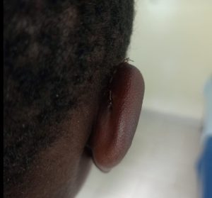
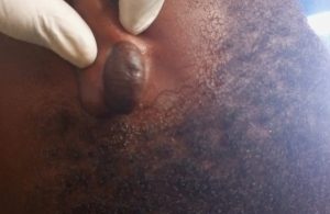
We report a case of a 14 year old female who presented with a history of a spontaneous and discomforting keloids on her right ear lobe. The patient was offered the treatment options of her choice and preferred surgical excision to intralesional triamcinolone.
Case Presentation
A 14 year old female black African, presented with a long standing history of a right ear lobe swelling that was noted since birth. The swelling has been progressing in size, intensely itchy and now painful. There was no any treatment attempted before. She reported that the swelling started spontaneously without any prior history of trauma.
On physical exam we noted a 4cm diameter dumbbell shaped, non tender stalked firm mobile mass occupying the posterior aspect of the right ear lobe. She was systemically stable with


normal vital signs. Her CBC parameters were all normal. A clinical diagnosis of spontaneous keloid was made. The patient was offered two treatment choices and the parents opted for surgical excision which was done under local anaesthesia using interrupted absorbable sutures. Currently she is under surveillance incase there may be recurrence.
DISCUSSION
This case study presented evidence that not all cases of keloid have a prior history of trauma.
Keloid represent aberrations in the fundamental processes of wound healing, in which there is an obvious imbalance between the anabolic and catabolic phases, in addition keloids seem to be a more sustained and aggressive fibrotic disorder. Evidence to date strongly suggests more prolonged inflammatory period, with immune cell infiltrate present in the scar tissue of keloids which may contribute to increased fibroblastic activity with greater and more sustained extracellular matrix deposition.
Keloids are more likely to be found in areas of the body that have high skin tension. From a histological perspective, keloids display an abundance of collagen settled in a haphazard way or whorls. As aforementioned, there is a higher incidence of keloids in people of Asian and African descent which positively correlates with increased melanin.
Spontaneous keloids are seen in those with novel X-linked disorders such as Dubowitz syndrome, Noonan syndrome, and Goeminne syndrome. There is also a positive correlation between keloids and known human leukocyte antigens such as HLA-B14, HLA-B21, HLA-BW16, HLA-BW35, HLA-DR5, and HLA-DQW3. Interestingly, evidence suggests that people with blood group A have a higher genetic association with the development of abnormal scars in general. We can clearly see that our patient being a black African is a risk factor for keloid in general however we could not ascertain wheather she had above genetic syndromes as their diagnosis would require expensive sophisticated testing modalities. The parents as well couldn’t remember whether the patient had micro trauma from other infections such as chicken pox.
CONCLUSION
Spontaneous keloid remains a rare entity and challenging to diagnose. Most of the reported cases were associated with certain syndromes which may raise the question of a genetic link between spontaneous keloids and certain mutations. In addition, it has also been reported in atopic persons and after the use of letrozol and isotretinoin therapy; however, a few reports denied any medical condition which put these cases in a doubtful situation, as there may have been a minor trauma that the patient was unware of, especially if it was only a single keloid.
REFERNCES
- Oluwasanmi JO: Keloids in the African. Clin Plast Surg 1974; 1: 179–195
- Moshref SS, Mufti ST. Keloid and hypertrophic scars: Comparative histopathological and immunohistochemical study. Med Sci. 2010;17:3–22
- Jain VK, Soundarya N, Rodrigues C, Shetty S. Bilateral tops like ear lobe keloid of unusual size: A case report and review of etiopathogenesis and treatment modalities. Int J Oral Maxillofacial Pathol. 2011;2:45–50
